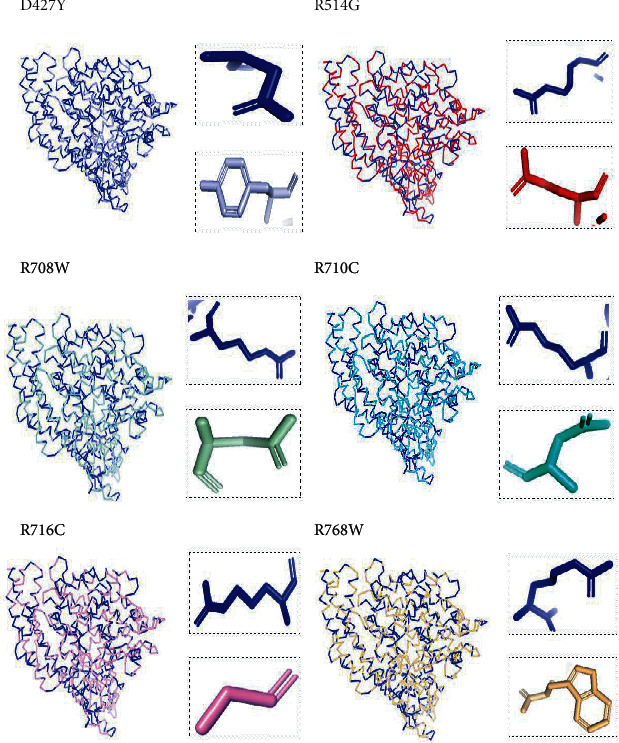
Figure 2.

Superimposed structure of native and mutant models of the ACE2 protein. The superimposed structure of native amino acid aspartic acid (blue color) with mutant amino acid tyrosine (light blue color) at position 427. The superimposed structure of native amino acid arginine (blue color) with mutant amino acid glycine (red color) at position 514. The superimposed structure of native amino acid arginine (blue color) with mutant amino acid tryptophan (pale green color) at position 708. The superimposed structure of native amino acid arginine (blue color) with mutant amino acid cysteine (green cyan color) at position 710. The superimposed structure of native amino acid arginine (blue color) with mutant amino acid cysteine (violet color) at position 716. The superimposed structure of native amino acid arginine (blue color) with mutant amino acid tryptophan (light orange color) at position 768.
