
FIG 2.
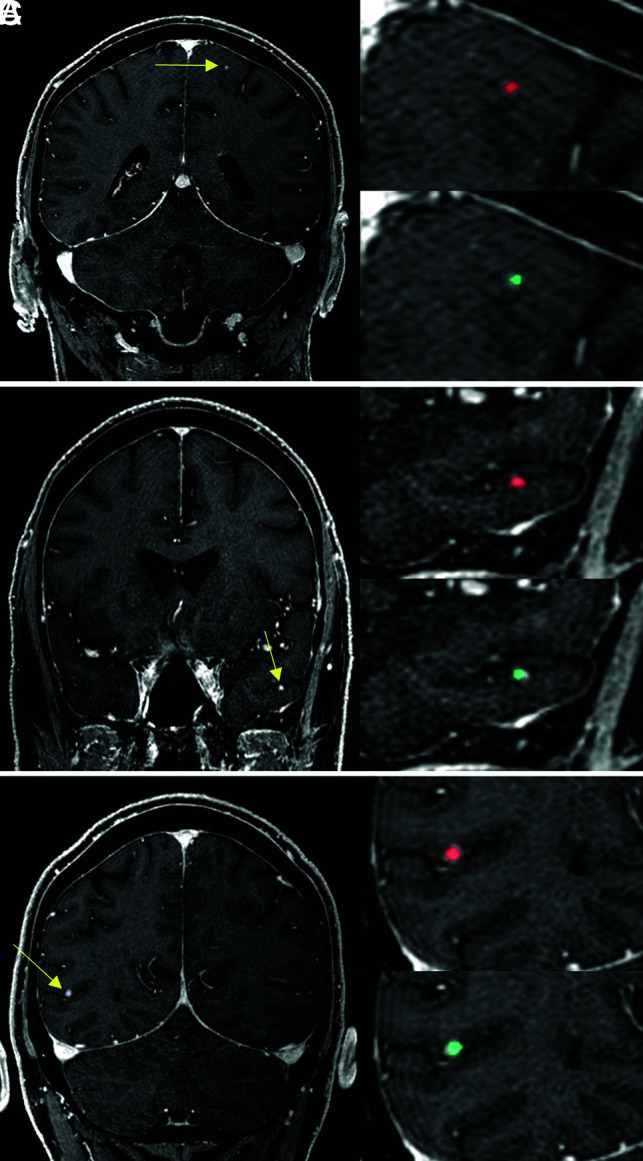
A 67-year-old male patient with malignant melanoma. The DLM (turquoise) detects the metastases (yellow arrows) of the left frontal lobe (A), the left temporal lobe (B), and the right parietal lobe (C) accurately and provides manual segmentations (red) comparable to segmentation performance.