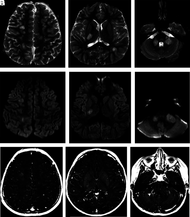
FIG 2.
Axial T2WI (A–C) demonstrates multiple large hyperintense oval lesions predominantly affecting the subcortical WM of the cerebral hemispheres, the posterior arm of the right internal capsule, and the infratentorial fossa structures, particularly in the middle cerebellar peduncles. All lesions concurrently demonstrate diffusion restriction observed in the diffusion sequence (D–F) and gadolinium enhancement in the postcontrast T1 sequence (G–I). Most lesions have an open-ring enhancement pattern, best characterized in the right middle cerebellar peduncle (arrowhead in I).

