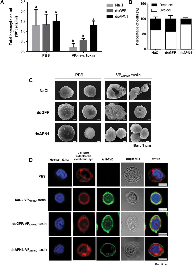
Fig 4. The effect of LvAPN1 silencing on shrimp hemocyte homeostasis.
(A) Effect of LvAPN1 silencing on the total hemocyte count after challenge with the partially purified VPAHPND toxins. Experimental groups were the same as those used in Fig 3B. PBS was used as a control. THC values for each treatment condition were derived from at least three shrimp. (B) Percentage of dead and viable hemoctyes in the LvAPN1 knockdown shrimp after partially purified VPAHPND toxins challenge was determined by trypan blue staining and observation under light microscopy. Asterisks indicate significant difference (P < 0.05). (C) The representative SEM micrograph showing morphology of LvAPN1 knockdown shrimp hemocytes after partially purified VPAHPND toxin challenge. Experimental groups were the same as those used in Fig 3B. PBS was used as a control. (D) Localization of the VPAHPND toxin on shrimp hemocyte by immunofluorescence. The VPAHPND hemocytic nuclei, cytoplasmic membrane and PirBvp toxin are visualized in blue (Hoechst 33342), red (CellBrite cytoplasmic membrane) and green (Alexa Fluor 488) colors, respectively. PBS-injected shrimp was used as a control. The scale bar corresponds to 5 μm. All experiments were done in triplicate.

