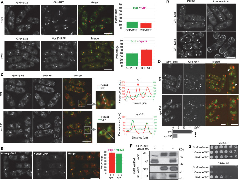
Fig 3. Stx8 cycles between the trans-Golgi network (TGN) and the prevaculolar endosome (PVE) in a retromer-dependent fashion.
(A) Colocalization between GFP-Stx8 and the TGN marker Cfr1-RFP (upper panels) or the PVE marker Vps27-RFP (lower panels). The experiments were performed two times, and a minimum of 400 GFP-Stx8 dots were scored in each experiment. The mean and standard deviation of the results are shown (right panels). For better accuracy, both the percentage of GFP-Stx8 dots that colocalized with Cfr1-RFP or Vps27-RFP dots and the percentage of Cfr1-RFP or Vps27-RFP dots that colocalized with GFP-Stx8 dots were determined. (B) Wild type (WT) cells bearing GFP-Syb1 or GFP-Stx8 were treated with dimethyl sulfoxide (DMSO; solvent) or latrunculin A for 20 minutes and photographed. (C) Distribution of GFP-Stx8 in WT and vps35Δ cells. GFP-Stx8 colocalization with FM4-64 is shown. Enlargement of representative vacuoles from the same strains are shown in the central panels. The right panels show line-scans of GFP and FM4-64 fluorescence intensities (a.u., arbitrary units) across the vacuoles, as indicated in the enlargements. (D) Colocalization between GFP-Stx8 and Cfr1-RFP in WT and vps35Δ strains. The right panels show enlarged cells. Arrowheads in the WT denote colocalization. The mean, standard deviation and statistical significance (Student’s t-test. Pvalue 0.0002) of the colocalization percentages from three independent experiments is shown in the lowest panel. (E) Colocalization between Cherry-Stx8 and Vps35-GFP. The mean and standard deviation of the results are shown (right panels). For better accuracy, both the percentage of Cherry-Stx8 dots that colocalized with Vps35-GFP dots and the percentage of Vps35-GFP dots that colocalized with Cherry-Stx8 dots were determined. Images are single planes captured with by confocal spinning disk microscopy (A, C, D and E) and with a DeltaVision system (B). Scale bar, 5 μm. (F) Stx8 and Vps35 coimmunoprecipitate. Cell extracts from stx8Δ vps28Δ strains carrying GFP-Stx8 and/or Vps35-HA were analyzed by western blot using anti-GFP or anti-HA monoclonal antibodies before (WCE, whole-cell extracts) or after immunoprecipitation (IP) with a monoclonal anti-GFP antibody. The asterisk denotes an unspecific band. (G) Serial dilutions of the Saccharomyces cerevisiae host strain AH109 strain transformed with empty plasmids (vector) and/or with plasmids expressing Stx8* (Stx8 lacking the TM helix) and/or a fusion Vps29-Vps35-Vps26 protein (CSC) were spotted on yeast nitrogen base medium without leucine and tryptophan (YNB-L-T) and without histidine (YNB-H) and incubated at 30°C for four days. Growth on YNB-H shows direct interaction between Stx8* and CSC.

