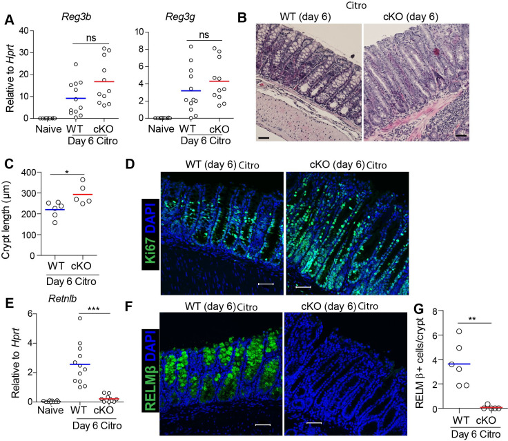
Fig 3. Intestinal-epithelial LSD1 is required for appropriate crypt hyperplasia and goblet cell responses to infection with C. rodentium.
(A) RT-qPCR for antimicrobials Reg3b and Reg3g in colon tissue of Naive (WT n = 10), and WT (n = 12) and cKO (n = 11) mice infected with C. rodentium for 6 days. (B&C) H&E staining and quantification of crypt length at distal colon in WT (n = 6) and cKO (n = 5) mice infected with C. rodentium for 6 days (scale bar = 50 μm). (D) Ki67 staining to measure proliferation in distal colon tissue of WT and cKO mice infected with C. rodentium for 6 days. (E) RT-qPCR for goblet-cell specific antimicrobials Retnlb in colon tissue of Naive (WT n = 10), WT (n = 12) and cKO (n = 11) mice infected with C. rodentium for 6 days. (F&G) Representative confocal images and quantification of RELMβ+ cells (green) in distal colon tissue of WT (n = 6) and cKO (n = 5) infected mice. DAPI (blue) was used as a nuclear counter stain. Scale bar = 50 μm. Unpaired two-tailed Student’s t test (A, C, E & G) was performed to observe significant differences among experimental groups. ns = not significant, * P ≤ 0.05, **, P < 0.01, *** P ≤ 0.001. Each dot represents one mouse and means are depicted.

