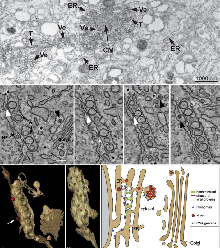
Fig. 2.
Flavivirus replication organelles derived from the ER. Top panel, TEM image of DENV replication organelles derived from the ER. T tubes, Ve virus-induced vesicles, CM convoluted membranes. Middle panel, continuous slices through an EM tomogram (~ 2 nm thick). White arrowhead, continuity of vesicle and ER membranes; black arrowhead, virus particles. Bottom panel, three-dimensional architecture of flavivirus replication organelles (left) and the model of flavivirus replication, assembly and release (right). These images are reproduced with permission from Ref. [7]

