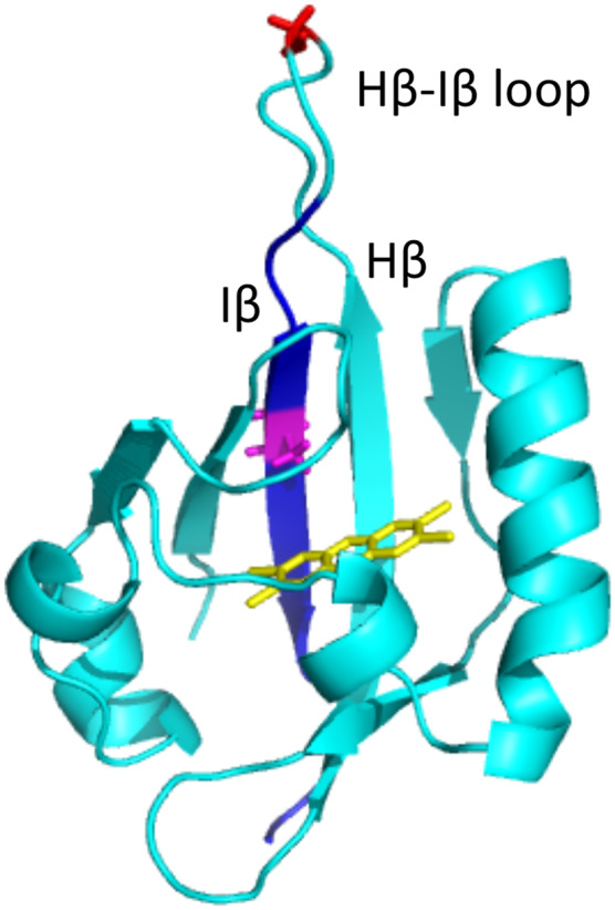
Figure 9.

Structural rendition of PAS-B from AHRb1 showing location of amino acids relevant to described mutants. The structure of the PAS-B domain modeled from AHRb1 sequence is shown in light blue. A TCDD molecule is shown in the ligand-binding pocket in yellow. Residue 375 (Alanine) is highlighted in purple. The Glycine residue at 368 mutated in the AHRNG367R model is highlighted in red andlocated in the hairpin loop. The residues changed in AHRTer383 as a result of the frame shift mutation are highlighted in dark blue.
