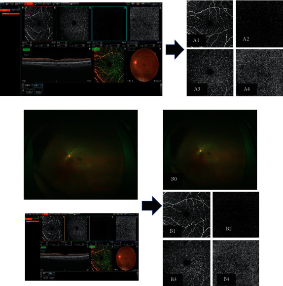
Figure 2.

Test 1 (no apparent diabetic retinopathy [NDR] or diabetic retinopathy [DR]) and test 2 (NDR or proliferative diabetic retinopathy [PDR]) were performed using the Optos (a), optical coherence tomography angiography (OCTA), Optos (b), Optos OCTA images (A1–A4; B0–B4). A1, B1: Superficial OCTA image; A2, B2: deep OCTA image; A3, B3: other retinal layer of the OCTA image; A4, B4: choriocapillaris layer of the OCTA image; B0: Optos image.
