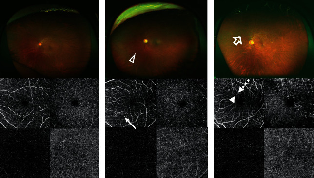
Figure 3.

Representative images of no apparent diabetic retinopathy (a), mild nonproliferative diabetic retinopathy (b), and proliferative diabetic retinopathy (c) obtained using ultra-wide-field (UWF) imaging and optical coherence tomography angiography (OCTA). The UWF image shows the hemorrhage (white triangle) and neovascularization (white arrow). The OCTA image shows microaneurysm (white long arrow), microvascular tortuosity (white dotted arrow), and capillary non-perfusion (white short arrow).
