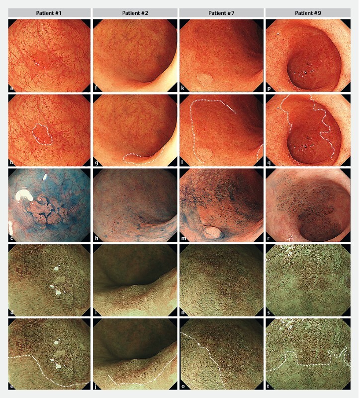
Fig. 1 .

Representative endoscopic images of flat-type dysplasia in UC patients. a, b, c, d, e Flat (Patient #1). f, g, h, i, j Flat (Patient #2). k, l, m, n, o Flat + sessile (Patient #7). p, q, r, s, t Flat lesion (Patient #9), according to the SCENIC consensus statement. These lesions were observed on white-light imaging (WLI) (the first and second row from the top), indigo‐carmine dye splaying (the third row) and magnifying narrow-band imaging (NBI) (the fourth and fifth row). Note that reddish areas on WLI were indicated by white dotted lines in the second row. The border of vascular increased areas on NBI was also demonstrated by white dotted lines in the fifth row.
