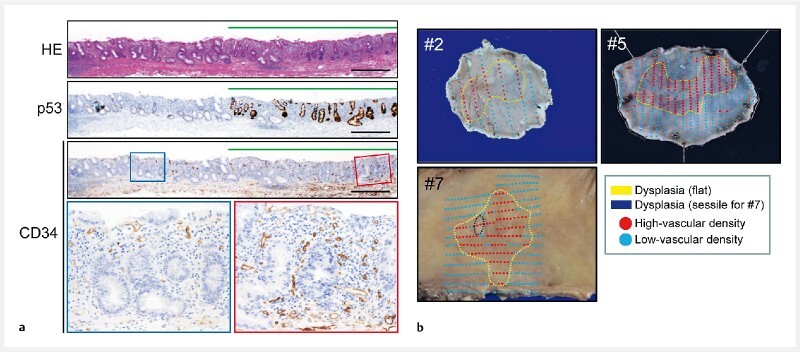
Fig. 2.

Representative histological and macroscopic images in flat-type dysplasia. a Representative histological images of flat-type high-grade dysplasia (HGD) (Case #7). The histological findings for the resected specimen: HE staining indicating a flat-type HGD lesion (upper) and p53 immunohistochemistry (middle) indicating overexpression of p53. Note that CD34-positive vessels in the mucosa were increased in the HGD lesions compared to in the adjacent non-neoplastic lesions by CD34 immunohistochemistry (lower). The rectangle dysplastic (red line) and non-neoplastic (blue line) areas are magnified below. Scale bars, 500 μm. b Representative macroscopic images of resected specimens of flat-type dysplasia showing mucosal vascular densities (Case #2, #5, and #7). Red and blue dots indicate high- and low-density of intramucosal vessels, respectively. The 75 th percentile was used to determine high- and low-density of intramucosal vessels as described in the “Patients and methods” section. Dysplastic areas confirmed by histological assessment were lined by yellow dotted lines. Case #7 contains a sessile lesion indicated by a black dotted line.
