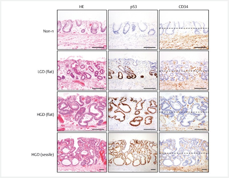
Fig. 3 .

Representative histological images of non-neoplastic, flat-type low-grade dysplasia, flat-type high-grade dysplasia and sessile high-grade dysplasia. The histological findings (Case #4): HE staining indicating non-neoplastic (Non-n), flat low-grade dysplasia (LGD), flat-type high-grade dysplasia (HGD) and sessile HGD lesions (left), p53 immunohistochemistry (middle), and CD34 staining indicating vessels in the mucosa (right). In CD34-immunostained sections, the luminal and basal half areas were divided by black dotted lines. Scale bars, 200 μm.
