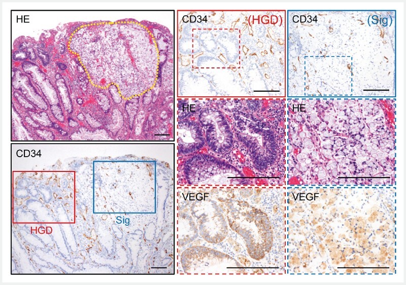
Fig. 6 .

Less vascular density/size in intramucosal signet-ring cell carcinoma areas. Representative histological images of colitis-associated tumor containing intramucosal high-grade dysplasia (HGD) and signet-ring cell carcinoma (Sig) components (Case #S1). HE staining (upper left) shows intramucosal HGD containing a Sig component which is lined by a yellow dotted line. CD34 staining indicates intramucosal vessels in these lesions (lower left). The rectangle HGD (red line) and Sig (blue line) areas in the CD34 staining are magnified to the upper right. Further magnified HE staining and VEGF staining were shown below. Scale bars, 200 μm.
