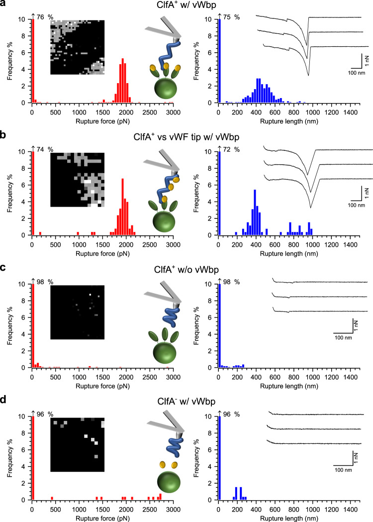
Fig. 2. vWbp augments ClfA-dependent binding of S. aureus to vWF and the ternary complex is very strong.
a S. aureus cells expressing ClfA at high levels and treated with recombinant vWbp probed with vWF-modified AFM tips. Data for a representative cell are shown. For more cells, see Supplementary Fig. 1. On the left are histograms of rupture forces with insets showing the respective adhesion maps (500 × 500 nm, 32 × 32 or 16 × 16 pixels, gray scale = 0–3 nN, each dot represents a binding event) and a cartoon in the top graph illustrates the experimental setup. Green ovals represent ClfA, golden spheres vWbp, and blue lines vWF. On the right are shown histograms of the rupture lengths with insets showing three representative retraction profiles. b Data for ClfA+ S. aureus cells (not treated with vWbp) probed with vWF-functionalized tips treated with vWbp. c Data for vWbp-untreated ClfA+ cells probed with vWF-modified tips. d Data for vWbp-treated ClfA− cells probed with vWF-modified tips.

