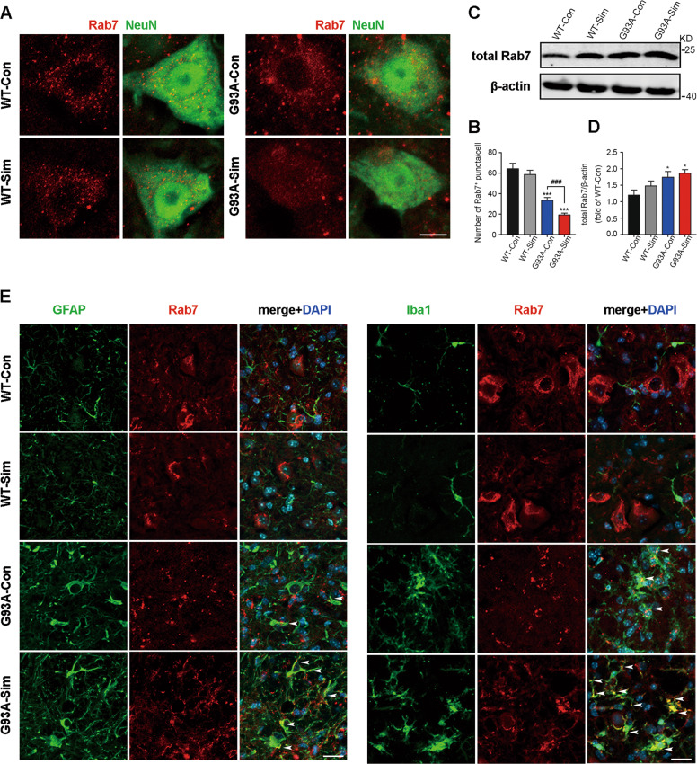
Fig. 5. Simvastatin blocked Rab7 localization to membranes in MNs from SOD1G93A mice.
A Immunofluorescence labeling of Rab7 (red) in NeuN-positive motoneurons (green) at 120 days (Scale bars, 10 μm). B Quantitative analysis of Rab7-positive puncta. C Western blot analysis of total Rab7 in the lumbar spinal cord at 120 days. D Quantification of total Rab7 levels. E Immunostaining of Rab7 (red) and GFAP (green), Rab7 (red) and Iba1 (green) in the lumbar spinal cord at 120 days (Scale bars, 20 μm). Nuclei were stained with DAPI (blue). White arrowheads show the portions of GFAP+/Rab7+ and Iba1+/Rab7+, respectively. (Data represent the mean ± SEM, n = 5 mice per group; statistical significance was assessed by one-way ANOVA or an unpaired t-test, *P ≤ 0.05, **P ≤ 0.01, ***P ≤ 0.001, #P ≤ 0.05, ##P ≤ 0.01, ###P ≤ 0.001).

