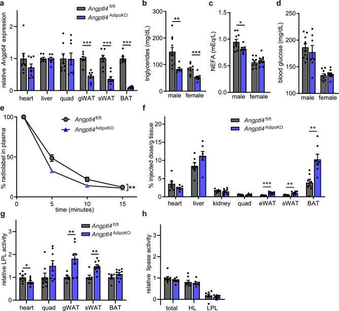
Figure 1.
Characterization of mice with adipocyte-specific deletion of Angptl4. (a) mRNA expression of Angptl4 in liver, heart, quadriceps muscle (quad), gonadal white adipose tissue (gWAT), subcutaneous white adipose tissue (sWAT), and brown adipose tissues (BAT) from 8–12 week old female Angptl4fl/fl (n = 8) and Angptl4AdipoKO (n = 8) mice following a 6 h fast (mean ± SEM). (b–d) Fasting plasma triglyceride (b), non-esterified fatty acid (c), and blood glucose (d) levels in 8–12 week male and female Angptl4fl/fl and Angptl4AdipoKO mice following a 6 h fast (mean ± SEM; n = 7–11). (e, f) Fasted (6 h) female Angptl4fl/fl (n = 8) and Angptl4AdipoKO (n = 7) mice were injected intravenously with 3H-triglyceride–containing chylomicrons. (e) Clearance of radiolabel from the plasma 1, 5, 10, and 15 min after injection. Points represent percentage of radiolabel remaining in the plasma at the indicated time points compared to the 1 min time point (mean ± SEM). (f) Uptake of radiolabel after 15 min (% injected dose/g tissue) into the indicated tissues (mean ± SEM). (g) Heart, quadricep muscle (quad), gonadal white adipose tissue (gWAT), subcutaneous adipose tissue (sWAT), and brown adipose (BAT) tissue from fasted (6 h) female Angptl4fl/fl and Angptl4AdipoKO mice were harvested and lipase activity was measured (n = 6–9/group). (h) Liver was harvested from fasted (6 h) female Angptl4fl/fl and Angptl4AdipoKO mice (n = 8/group). Lipase activity was measured in the presence or absence of 1 M-NaCl to distinguish between hepatic and lipoprotein lipase. Bars show relative lipase activity in each tissue normalized to Angptl4fl/fl (mean ± SEM). *p < 0.05, **p < 0.01, ***p < 0.001 by t-test analysis (panels a–c, f, g) or repeated measures ANOVA (panel e).

