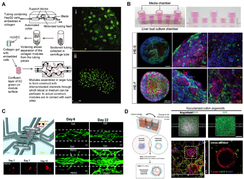
Fig. 3.
Modeling for vascularized organoids-on-a-chip. a The approach for collagen-HepG2 modules to build vascularized organoids. The HepG2 cells collagen cylinders covered with human umbilical vein endothelial cells (HUVECs) were cultured in the flow circuit enabling perfusion with medium or blood to deliver nutrients to the assembly. Confocal microscopy image of (i) vascular endothelial (VE)-cadherin staining of HUVEC layer on the construct and (ii) prelabeled viable HepG2 cells [212]. b 3D vascularized hepatic organoids in a rocker-actuated microfluidic system. Confocal images for CD31 (green) and albumin (ALB; red) of the liver organoids consisting of induced hepatic cells, HUVECs, and a decellularized liver extracellular matrix cultured under each condition demonstrate increased albumin expression and vascular network of the liver organoids when cultured with media flow: under static conditions (iHE-S) and dynamic conditions induced by the microfluidic system (iHE-F). Scale bars, 500 μm (white), 50 μm (yellow) [213]. c A microfluidic device to coculture patient-derived tumor organoids (PDTO) and a perfusable microvascular networks. Fluorescently tagged PDTOs (red; scale bar, 50 μm) cultured in the microfluidic chip grow in the pre-vascularized system. The vascular network (green; scale bar, 100 μm) cocultured with PDTOs show highly angiogenic features [214]. d A microfluidic platform to engineer vascularized colon organoids. Confocal microscopy images of vascularized colon organoids for F‐actin (red), DAPI (blue), and GFP‐endothelial cells (green). [215]

