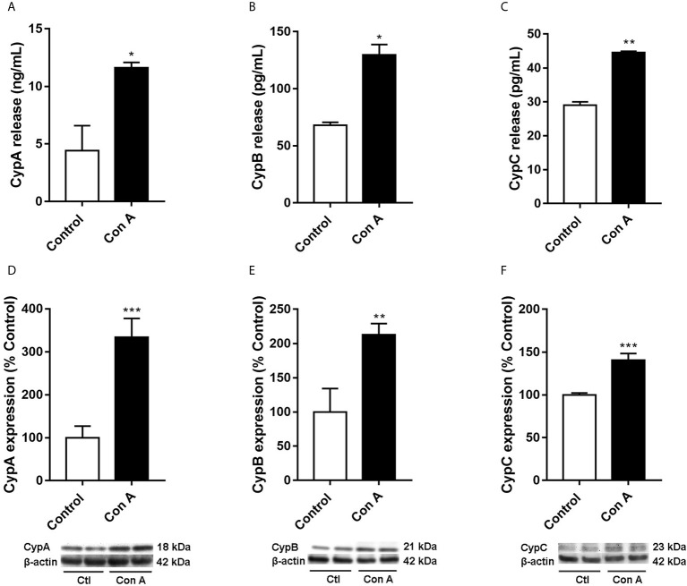
Figure 2.
Intra and extracellular CypA, B, and C levels in human T lymphocytes stimulated with Con A Cells were treated with Con A (50 μg/mL) for 48 h. CypA (A), CypB (B), and CypC (C) levels were measured by ELISA in the medium of T lymphocytes. The intracellular expression of CypA (D), CypB (E), and CypC (F) was measured by western blot in cytosolic lysates of T lymphocytes. Band intensity was normalized by β-actin. Data are the result of average ± SEM (n = 3). The values are shown as the difference between cells treated with Con A and untreated cells, *p < 0.05, *p < 0.01 and ***p < 0.001. One-way ANOVA test with Dunnet’s post hoc analysis and Student´s t-test were used for statistical analysis.

