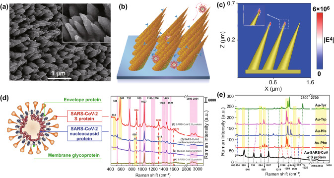
Fig. 2.
Morphology analysis of gold-nanoneedles array and SERS spectra of viral protein. a SEM images of Au nanoneedles array fabricated by Ar+ ions irradiation at a tilted angle of 45° on an Au film of 500 nm in thickness. b Schematics of “virus-traps” nanoforest composed of tilted gold-nanoneedles array. c Calculated intensity distribution (|E|2) at 785 nm for a tilted Au nanoneedle array with the polarization of the incident laser along the x-axis. d Structure schematics of SARS-CoV-2 (left), and SERS spectra (right) of SARS-CoV-2 S protein and nucleocapsid protein, SARS-CoV S protein, and Human ACE2 protein at 100 nM level. e Calculated static Raman spectra of 4 types of main individual amino acids Tyr, Trp, His, Phe encoded in SARS-CoV-2 S on Au cluster

