Figure 5.

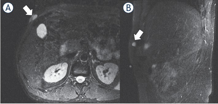
1.5-T MRI of the abdomen (A) contrast-enhanced T1 axial, (B) contrast-enhanced T1 sagittal) of a 41-year-old patient. The low-grade sarcoma in the right abdominal wall (white arrow) presents with a ovoid/nodular configuration.

1.5-T MRI of the abdomen (A) contrast-enhanced T1 axial, (B) contrast-enhanced T1 sagittal) of a 41-year-old patient. The low-grade sarcoma in the right abdominal wall (white arrow) presents with a ovoid/nodular configuration.