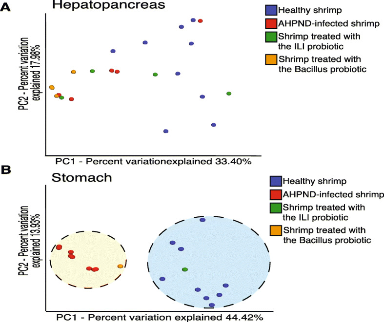
Fig. 3.

Beta diversity analysis of microbiota from hepatopancreas and stomach samples. a A PCoA graphical representation based on Bray–Curtis similarity does not show a clear grouping pattern between different treatments for hepatopancreas samples in the two main axes explaining over 50% of the variation. b PCoA plot showing a clear grouping pattern that separates stomachs of infected from healthy samples. Variations between groups highlight the clustering of the ILI probiotic with the healthy shrimp, while the Bacillus probiotic seems closer to the infected individuals. Notice that all samples were ran in the same PCoA analysis but are separated by organ for visualization purposes. Each colored circle corresponds to a type of sample; healthy individuals (blue), infected individuals (red), with ILI probiotic (green) and Bacillus probiotic (orange). Probiotic treatments in panel B represent a pool of 5 stomachs processed together
