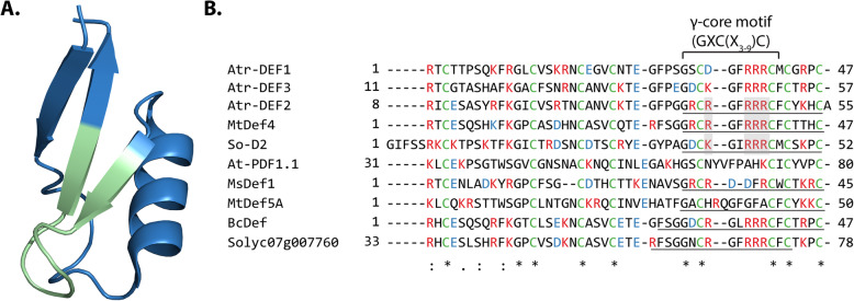
Fig. 1.
(a) 3D structure of Medicago truncatula defensin MtDef4 (PDB: 2LR3, figure generated in Pymol) [15] with its γ-core motif (GXCX3-9C, where Xn is the number of residues between cysteines) highlighted in green. (b) Alignment of Atr-DEF2 (predicted mature sequence) with other plant defensins including known sequence of Atr-DEF1 [16] and predicted mature sequence of Atr-DEF3. Underlined regions have been synthesized as γ-core motif analogs [8, 17, 18]. Cysteines forming disulfide bonds are green, basic residues are red, and acidic residues are blue. Conserved basic residues of membrane lytic γ-core motif analogs are shaded grey. Asterisks note fully conserved residues, two dots note positions with highly similar residues, and single dots note positions with weakly similar residues

