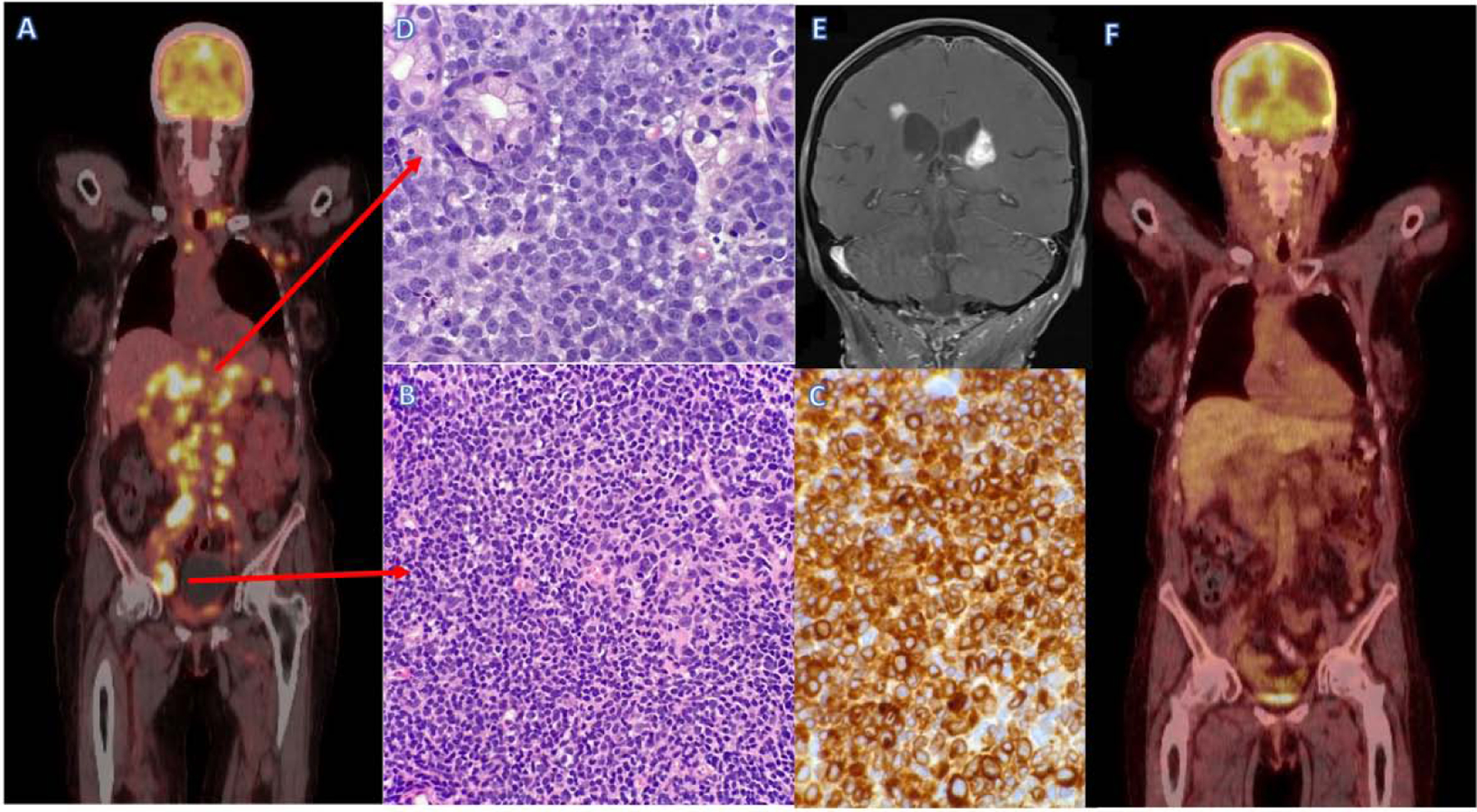
Figure 2: Tumor heterogeneity and response to treatment.

A; Pre-treatment PET/CT scan in newly diagnosed lymphoma demonstrates advanced stage disease.
B; Inguinal lymph node biopsy demonstrating low-grade follicular NHL, H&E stain (x40 magnification).
C; Inguinal lymph node biopsy demonstrating low-grade follicular NHL, BCL-2 IHC stain (x60 magnification).
D; Gastric ulcer biopsy demonstrating high grade B-cell lymphoma DLBCL, H&E stain (x60 magnification).
E; End of treatment PET/CT scan demonstrates near complete remission.
F; MRI scan after NHL treatment demonstrates new peri-ventricular white matter lesions suggestive of NHL relapse.
