Figure 8.

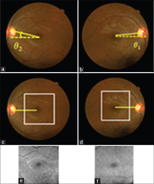
(a and b) Fundus images before alignment, (c and d) Fundus image after alignment with the specified area of optical coherence tomography projection, (e and f) corresponding optical coherence tomography projections

(a and b) Fundus images before alignment, (c and d) Fundus image after alignment with the specified area of optical coherence tomography projection, (e and f) corresponding optical coherence tomography projections