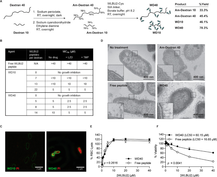
Figure 3.
Potentiator candidate WD40 selectively disrupts the bacterial membrane to allow small molecule influx into the cytoplasm. (A) Synthetic scheme for potentiator candidates—WD10 and WD40. (B) Table showing MIC90 values for WD10 and WD40 containing different number of WLBU2 peptides (n = 3). MIC90 values were determined in a microdilution assay with P. aeruginosa (strain PA14) in which different dilutions of potentiator candidates were combined with a fixed drug concentration—5 μM free linezolid (LZD) or 5 μM trimethoprim (TMP). (C) Fluorescent images acquired using a super-resolution microscope to visualize PI influx in PA14 with WD10 or WD40 treatment. Images are representative of observations from two independent imaging experiments. (D) Transmission electron microscopy (TEM) images of the PA14 untreated control and PA14 after a 5 min incubation with Am-Dextran 40 (1 μM), free WLBU2 peptide (5 μM), or WD40 (5 μM). Images show disruption of the bacterial membrane by free peptide and WD40. (E) Comparison of the concentration-dependent hemolytic activity of WD40 versus free WLBU2 peptide (n = 3). No significant difference was observed between the two treatments. Repeated measures ANOVA was used to determine the p-value. (F) Comparison of the concentration-dependent cytotoxicity of WD40 versus free WLBU2 peptide in HEK293T cells (n = 3). WD40 is significantly less toxic than free WLBU2 peptide. Repeated measured ANOVA was used to determine p-value. Lethal concentration for 50% cells (LC50) was determined using nonlinear regression to fit a dose–response curve.

