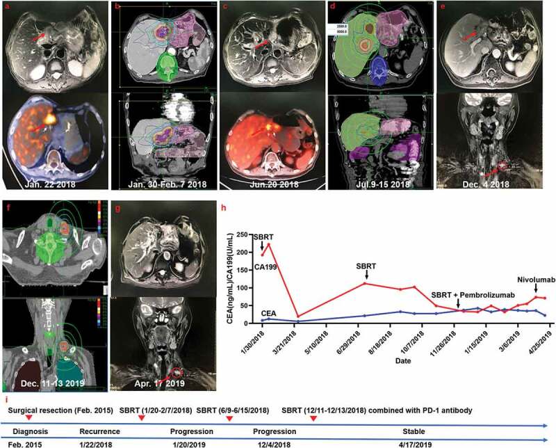
Figure 3.

Summary of imaging scans and timeline of therapy and disease status for Case 3
(a) The recurrent status after surgical resection. (b) The patient then received stereotactic body radiotherapy (SBRT) for the recurrent lesion. (c) MRI scans 4 months after RT revealed progressive enlargement of the recurrent lesion and growth of a new hepatic hilar metastasis. (d) The second SBRT for the recurrent lesion and the new hepatic hilar metastasis. (e) Further disease progression with a left supraclavicular lymph node metastasis detectable in December 2019. (f) The third SBRT for the left supraclavicular lymph node metastasis. (g) The disease stabilized after anti-PD-1 immunotherapy administration. (h) Serum carbohydrate antigen 199 (CA199) and carcinoembryonic antigen (CEA) levels with respect to the treatment timeline. (i) Timeline showing therapy and disease status throughout the disease course.
