
Fig. 2.
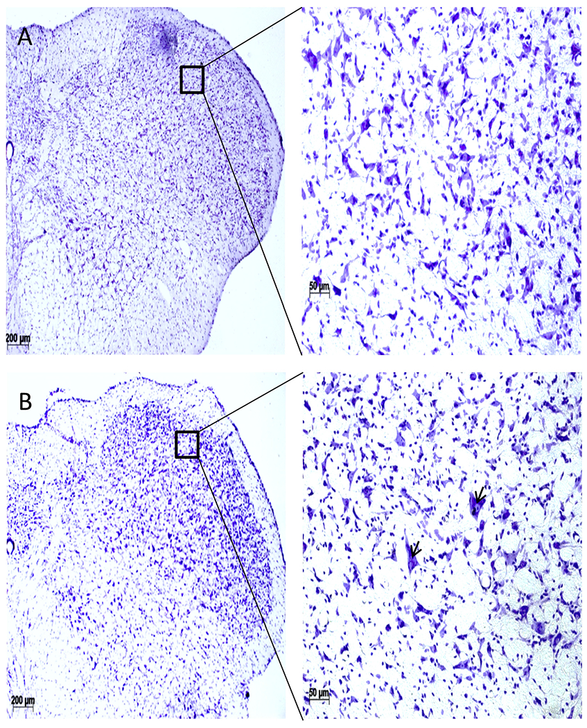
Cresyl-violet staining of the 5th cranial nucleus (SP5C), coronal sections (A, Normal; B, TBI). TBI SP5C area (B) did not show any detectable changes in the cellular morphology except appearance of a few largers cells (arrowhead). Scale bar = 100 μm.