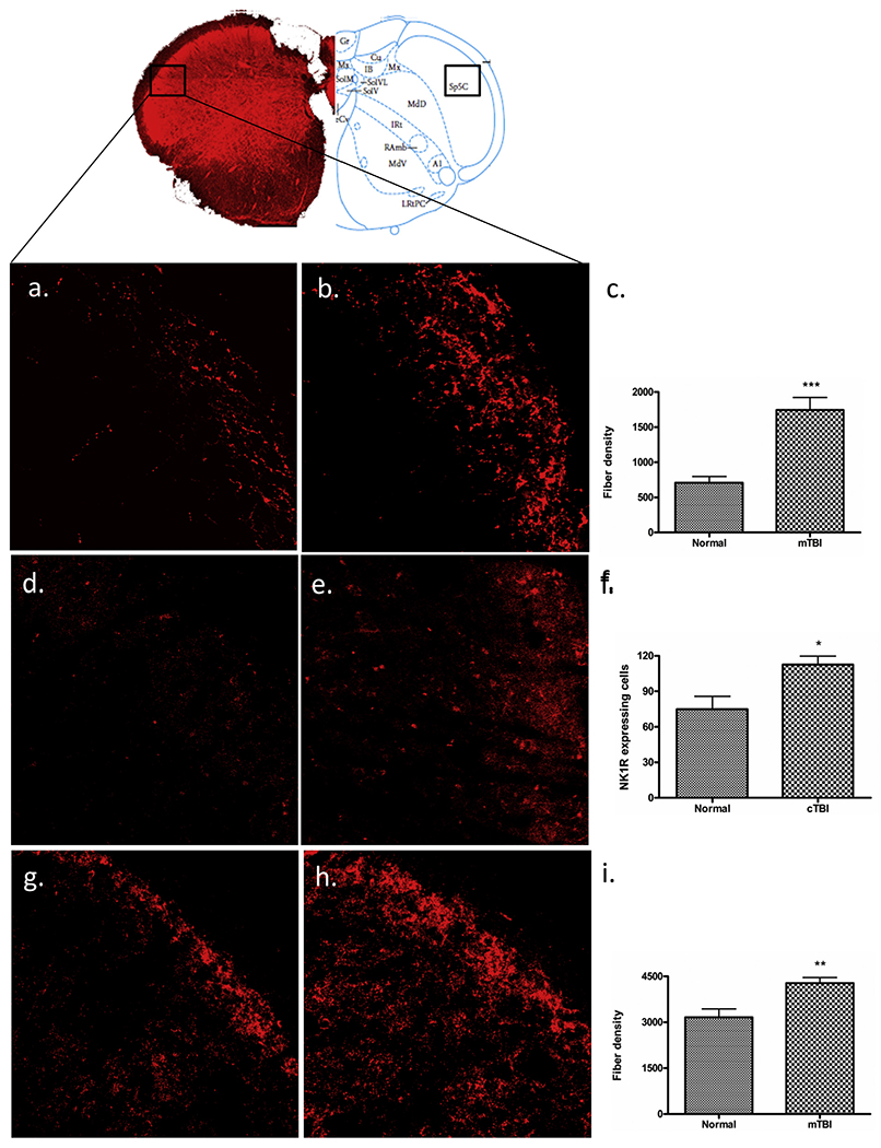
Fig. 5.

Immunofluorescent labelling of SP5C. Mild-TBI altered the immunoreactive expression of 5-HT (b), NK1R (e), and GAD67 (h) in the 5th cranial nerve nucleus caudalis (B: −15.0) according to the Paxinos atlas compare to the corresponding areas of control animals (a, d, g). Scale bar = 50 μm. A quantitative evaluation of fiber density for 5-HT (c), GAD67 (i) and NK1R expressing cells (f) showed significant elevation following m-TBI. “*”, P < 0.05, “**”, p < 0.005, and “***”, indicates P < 0.0005.
