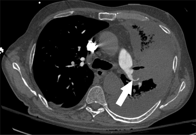
Figure 1d:

Examples of study- and image-level labels. (a) Central Pulmonary Embolism (PE): saddle embolus within the main, right, and left pulmonary arteries (arrow). (b) Right-sided PE and Left-sided PE: pulmonary emboli within the right interlobar, right lower lobe (arrow), and lingular pulmonary arteries (arrowhead). (c) Chronic PE: nonocclusive intraluminal web within the right lower lobe pulmonary artery (arrow). (d) True Filling Defect not PE: left lung malignancy invading the left main pulmonary artery. (e) Flow Artifact: an apparent filling defect within the left pulmonary artery, which is due to laminar flow of contrast media rather than PE (arrow). (f) RV/LV Ratio: < 1: normal RV (red line) to LV (blue line) ratio. (g) RV/LV Ratio: ≥ 1: evidence of right heart strain characterized by an elevated RV/LV ratio. (h) QA-motion: impaired image quality at the lung bases due to respiratory motion (arrows). (i) QA-contrast: insufficient opacification of the pulmonary arterial tree (arrow) to allow for the assessment of PE. LV = left ventricle, QA = quality assurance RV = right ventricle.
