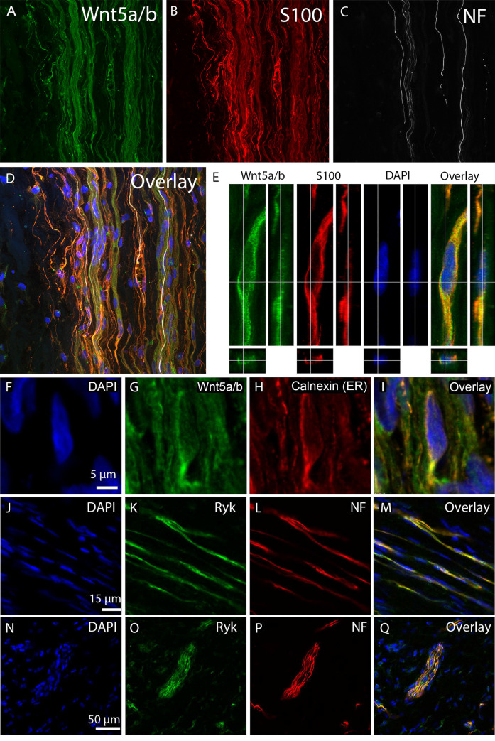
Fig 2. Wnt5a/b is expressed by Schwann cells in NIC.
A-E, Immunohistochemistry for Wnt5a/b on human NIC tissue sections. Wnt5a/b (A, green) is expressed by S100 (B, red) positive Schwann cells in the NIC. Neurofilament (NF) positive axons (C, gray) that traverse the NIC are closely associated with the Schwann cells (D, overlay (Including nuclear marker DAPI, blue)). E, High magnification with orthogonal view of an immunohistological staining for Wnt5a/b (green), S100 (red) and DAPI (blue) shows the presence of Wnt5a/b protein in a S100 positive Schwann cell. F-I, Strong Wnt5a/b (G, green) expression is visible in intracellular perinuclear structures (blue in F). Co-staining with the ER-marker calnexin (H, red) reveals that Wnt5a/b is expressed in the ER (I, overlay). J-M, N-Q, Cellular localisation of Ryk in NIC. Co-staining for DAPI, Ryk and Neurofilament reveals that Ryk expression (green in K, O) is observed on individual neurofilament positive axons (L, red) and in axon bundles (P, green) that traverse the human NIC (M, Q, overlay). Panel N to Q also show that Ryk is also weakly expressed around the nuclei (blue in N) of cells outside axon bundles in the NIC (Q, overlay).

