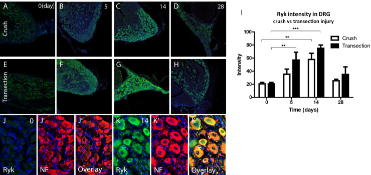
Fig 9. Ryk protein expression is increased in axotomized DRG neurons.
A-H, Immunohistochemical staining of Ryk (green) in L5 DRG following sciatic nerve crush (top row) or transection (bottom row) ranging from control (0 day) to 28 days p.i. (right column). Ryk staining intensity is low in control DRG neurons (A, E). Ryk staining intensity increased over time, and peaks at 14d p.i. (C, G) and returns towards baseline at 28d p.i. (D, H). I, Quantitative analysis of Ryk expression in DRG following sciatic nerve lesion (* p<0.05, ** p<0.01, *** p<0.001, error bars represent SEM). J, K, High magnification of DRG tissue sections. Immunohistochemistry for RYK (green) and NF (red) shows that control neurons (J) express very low amounts of RYK (J’). The increase in RYK expression at 14 days following nerve transection (K) is mainly due to an increase in neuronal (NF, K’) expression of RYK (Overlays in J” and K”).

