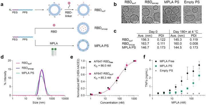
Figure 1 |. RBD and MPLA are formulated into stable, biologically active polymersomes (PS).
a, Schematic of formulation of PS. RBD was conjugated to the surface (RBDsurf) or encapsulated inside (RBDencap) of PS, and MPLA was encapsulated in the vesicle membrane (MPLA PS) due to its hydrophobicity. b, Representative cryo-electron microscopy images of PS, depicting vesicle structure. Scale = 50 nm. c, Size and polydispersity index (PDI) from dynamic light scattering (DLS) measurements of PS upon formulation and after > 6 months at 4 °C. d, Representative DLS curves of PS. e, Normalized mean fluorescence intensity (MFI) of AF647 conjugated to free RBD or RBDsurf by flow cytometry showing concentration-dependent binding to HEK-293 cells that express human ACE-2 (HEK-hACE2). Nonlinear regression was used to model data assuming specific binding to one site to determine equilibrium dissociation constants. f, Dose-dependent secretion of TNFα from cultured murine bone marrow-derived dendritic cells (BMDCs) stimulated by free MPLA, MPLA PS, or empty PS. Data represent mean ± SD for n = 2 (e) or 3 (f) replicates.

