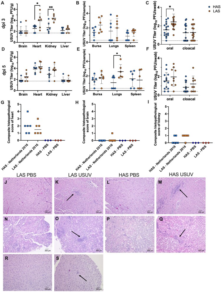
Figure 5.
Evidence of viral dissemination, shedding, and histopathology in tissues of HAS and LAS chickens inoculated with USUV. (A) Viral titer in tissues collected on dpi 3. (B) Viral titer in tissues collected on dpi 3. (C) Viral titer in oral and cloacal swabs collected on dpi 3. (D) Viral titer in tissues collected on dpi 5. (E) Viral titer in tissues collected on dpi 5. (F) Viral titer in oral and cloacal swabs collected on dpi 5. Circles represent individual samples; lines represent mean; error bars represent standard deviation. The limit of detection is indicated by dashed line. *p < 0.05, **p < 0.01. (G) Composite score of heart tissue; line represents median. (H) Composite score of brain tissue; line represents median. (I) Composite score of kidney tissue; line represents median. (J) Representative image of heart tissue collected on dpi 5 from LAS chickens inoculated with PBS, with no inflammation (H&E stain). (K) Representative image of heart tissue collected on dpi 5 from LAS chickens inoculated with USUV, with inflammatory lesions and presence of heterophils and lymphocytes (arrow) (H&E stain). (L) Representative image of heart tissue collected on dpi 5 from HAS chickens inoculated with PBS, with no inflammation (H&E stain). (M) Representative image of heart tissue collected on dpi 5 from HAS chickens inoculated with USUV, with inflammatory lesions and presence of heterophils and lymphocytes (arrow) (H&E stain). (N) Representative image of kidney tissue collected on dpi 5 from LAS chickens inoculated with PBS, with no inflammation (H&E stain). (O) Representative image of kidney tissue collected on dpi 5 from LAS chickens inoculated with USUV, with heterophilic inflammatory foci (arrow) (H&E stain). (P) Representative image of kidney tissue collected on dpi 5 from HAS chickens inoculated with PBS, with no inflammation (H&E stain). (Q)Representative image of kidney tissue collected on dpi 5 from HAS chickens inoculated with USUV, with heterophilic inflammatory foci (arrow) (H&E stain). (R) Representative image of brain tissue collected on dpi 5 from LAS chickens inoculated with PBS, with no inflammation (H&E stain). (S) Image of brain tissue collected on dpi 5 from LAS chicken inoculated with USUV, with proliferation of glial cells in the neuropil (arrow) (H&E stain, scale bars = 200 µm).

