Image 3.

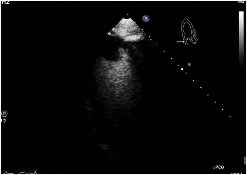
ECHO: Image obtained after administration of contrast agent, showing spherical filling defect noted in apex, consistent with apical thrombus

ECHO: Image obtained after administration of contrast agent, showing spherical filling defect noted in apex, consistent with apical thrombus