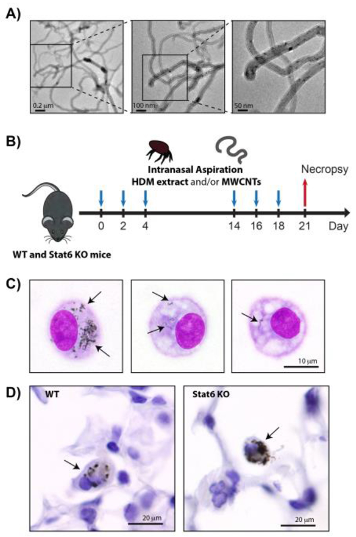
Figure 1.

A) TEM images of the MWCNTs used in this study. B) Illustration of exposure protocol timeline. Wild type (WT) and Stat6−/− mice were exposed repeatedly to HDM extract and/or MWCNTs by intranasal aspiration at days 0, 2 and 4, followed another round of repeated intranasal aspiration exposure at days 14, 16 and 18. Necropsy was performed at day 21 to collect BALF, lung tissue and serum. See the Methods section for details. C) Oil immersion (1000x) images of alveolar macrophages isolated by Cytospin centrifugation from the BALF of WT mice 21 days after exposure to MWCNTs using the protocol illustrated in panel B. Arrows indicate MWCNTs in the cytoplasm. D) Oil immersion (1000x) images of MWCNTs in the lung tissue of WT and Stat6 mice in situ. Arrows indicate macrophages with MWCNT inclusions.
