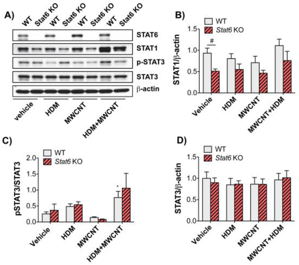
Figure 7. STAT protein levels and activation in lung tissue from WT and Stat6 KO mice after treatment with HDM extract in the absence or presence of MWCNTs.

Protein lysates of right lung lobe tissue collected from mice at necropsy were assayed by Western blotting for STAT proteins and β-actin. A) Representative Western blots of lung tissue from WT or Stcit6 KO mice exposed to vehicle, HDM, MWCNTs, or HDM and MWCNTs. B)Densitometry of STAT1 signal normalized against the β-actin signal. #P < 0.05 between genotypes, one-way ANOVA. N=5 animals per group. C)Densitometry of p-STAT3 normalized against total STAT3. *P < 0.05 compared to vehicle, Mann-Whitney test. D)Densitometry of total STAT3 normalized against β-actin. N=4 to 6 animals per group.
