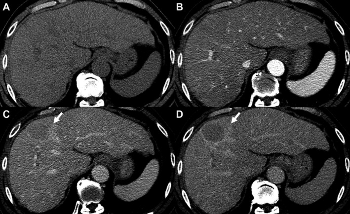
FIG 1.

A 62‐year‐old patient with hepatitis C cirrhosis. Multiphasic CT of the liver demonstrates a 42‐mm observation (arrows) with no APHE relative to precontrast images (A and B). This observation demonstrates nonperipheral washout on portovenous phase (C) and an enhancing capsule on delayed phase (D). This lesion is proved pathologically to be HCC.
