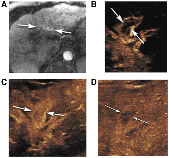
FIG 1.

CEUS LR‐TIV in a 61‐year‐old man with alcoholic cirrhosis. (A) An axial arterial phase contrast‐enhanced T1‐weighted fat‐suppressed MR image shows arterial hyperenhancement in the left portal vein area (arrows) suspicious for TIV. Delayed MR images were nondiagnostic due to motion artifact. CEUS images of the left portal vein show curvilinear arterial enhancement (arrows in B, 8 seconds) with complete enhancement later in the arterial phase (arrows in C, 18 seconds) and washout in the delayed phase (arrows in D, 3 minutes 45 seconds), diagnostic of TIV.
