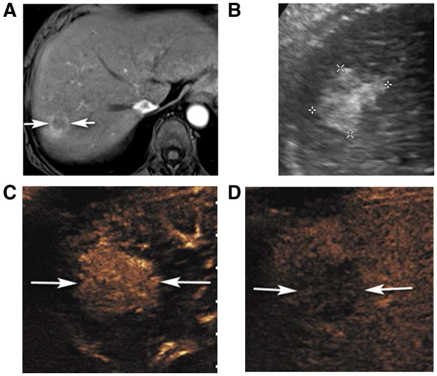
FIG 3.

CEUS LR‐5 in a 67‐year‐old woman with hepatitis C cirrhosis and CT/MRI LR‐M observation. (A) An axial arterial phase contrast‐enhanced T1‐weighted fat‐suppressed MR image shows a rim enhancing observation (arrows). (B) Corresponding ultrasound image shows an irregularly marginated solid, echogenic nodule (between calipers). CEUS shows diffuse, heterogeneous APHE of the nodule (arrows in C, 23 seconds) with definite washout on late phase (arrows in D, 4 minutes 7 seconds) enabling characterization as CEUS LR‐5. Biopsy showed HCC.
