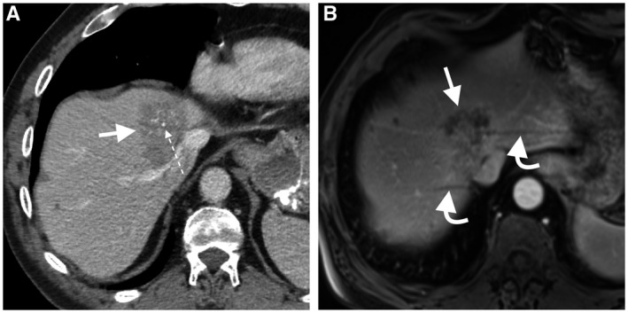
FIG 2.

A 65‐year‐old woman with hepatic metastasis from mucinous colorectal cancer. Central calcification (dotted arrow) within a hypoattenuating liver mass is seen only on portal venous phase CT image (A). The mass is heterogeneous hypoenhancing on the axial dynamic postcontrast portal venous‐phase MRI (B), but calcification is not seen. Also, note the respiratory motion artifacts on MRI (curved arrows), a recognized limitation of MRI in the assessment of liver lesion close to the hepatic dome.
