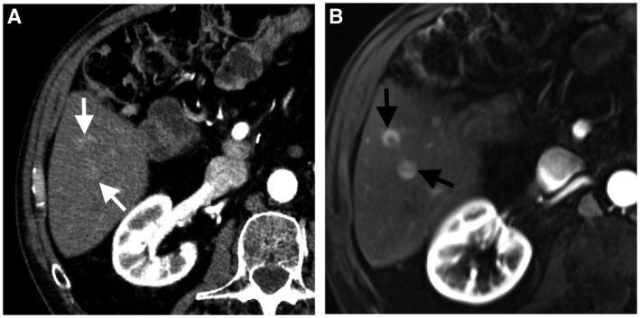
FIG 3.

A 43‐year‐old man with hepatic metastasis from pancreatic neuroendocrine tumor. Axial arterial‐phase CT image (A) shows two subtle arterial‐phase hyperenhancing metastatic nodules in hepatic segment 6 (white arrows). Axial arterial‐phase dynamic postcontrast MRI (B) shows improved visibility of subtle metastases (black arrows).
