. 2021 Mar 31;10:e65699. doi: 10.7554/eLife.65699

Figure 4. Chemical cross-linking and mass spectrometry reveals THO–Gbp2 interactions.

(A) Circular plot showing the cross-linking sites with EDC cross-linker. Each THOGbp2 complex subunit is represented as a colored segment with the amino acid residues indicated. Intermolecular cross-links are mapped inside the circle and the intramolecular cross-links are mapped outside the circle. The cross-links between Tho2 and Gbp2 are colored in orange. (B) Schematics of the arrangement of the Tho2–CTD, which contains a ‘bridge’ connecting two THO molecules, followed by a structured segment and a flexible tail (residues ~1400–1597). The EDC cross-links between the structured Tho2–CTD fragment and Hpr1 (E297, D434, and K462) as well as Mft1 (D129) are indicated by yellow lines. The DSS cross-link between Tho2–CTD and Hpr1–K467 is indicated by a purple line. (C) Schematics of the THO–Gbp2 interactions (left) and the identified cross-linking sites between Tho2–CTD and Gbp2 RRM domains. (D) In vitro GST-pull downs show that Gbp2 binds to the THO•Sub2 complex.

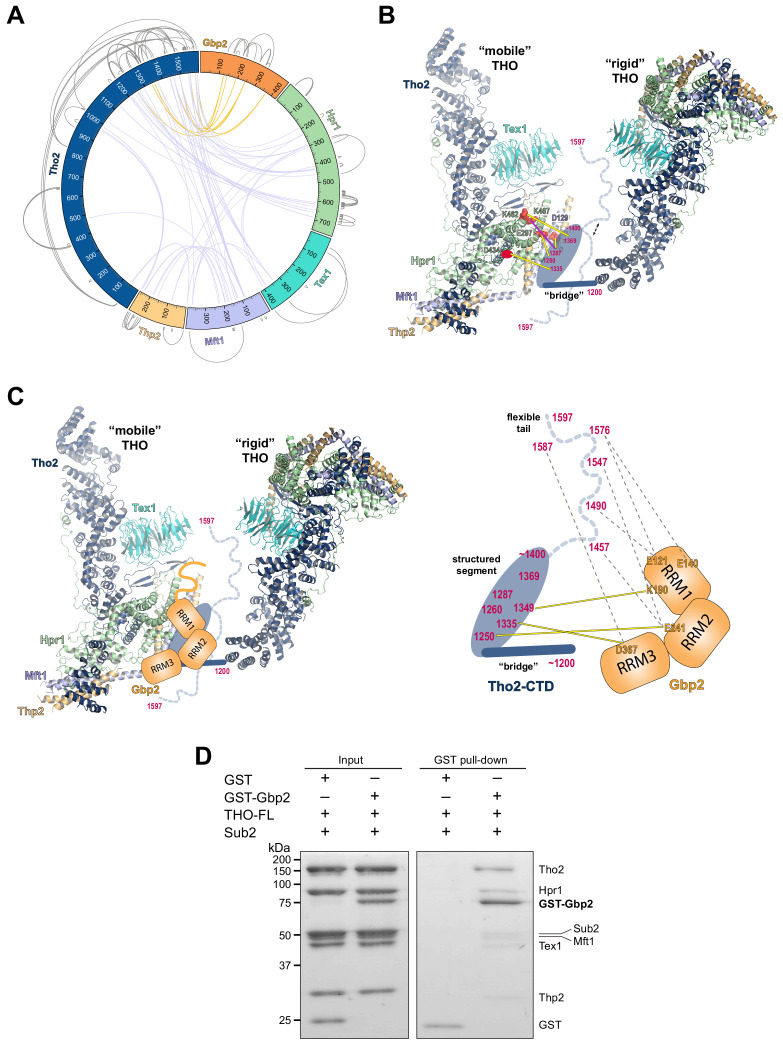
Figure 4.

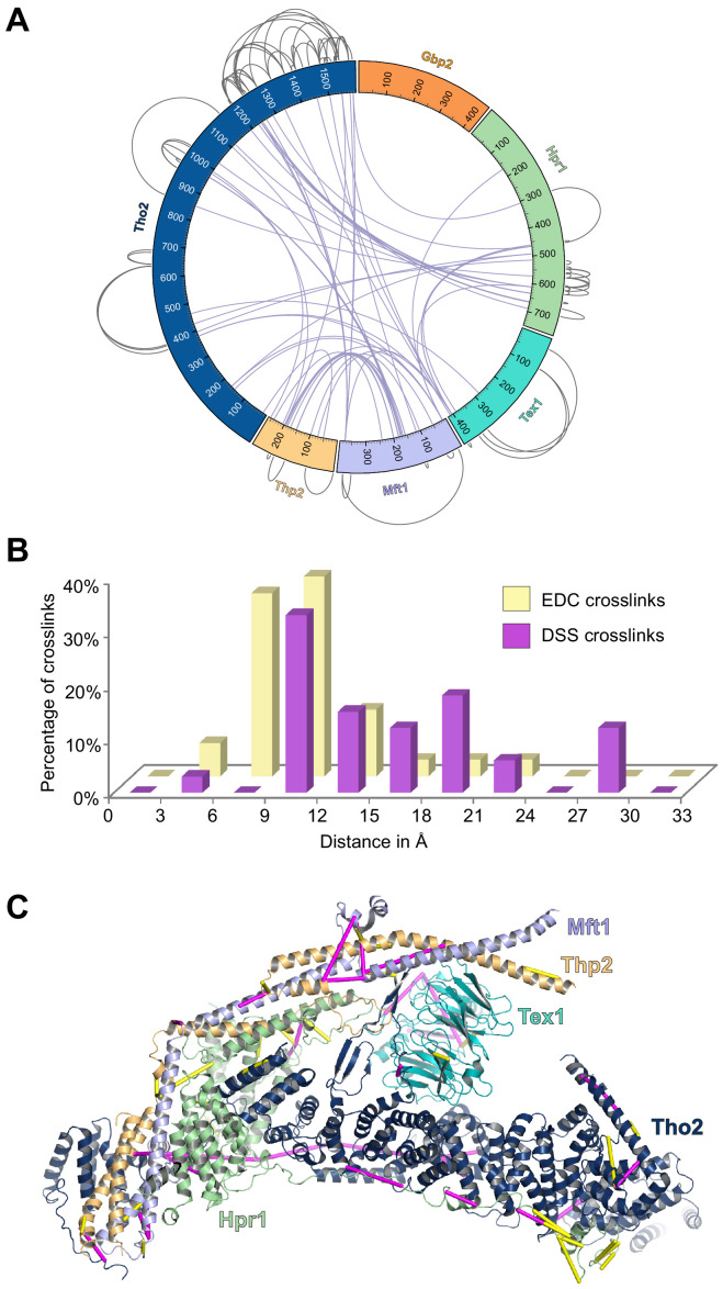
Figure 4—figure supplement 1. Analyses of the XL-MS data.

Figure 4—figure supplement 1.

(A) Circular plot showing the cross-linking sites with DSS cross-linker. (B) Distance distribution of the cross-links. We mapped the Cα–Cα distances between cross-linked residues onto the dimeric THO structure (PDB ID 7AQO). 91% of the EDC cross-links and 100% of the DSS cross-links that can be mapped to the structure fall within the expected threshold of 17 Å and 30 Å. (C) EDC (yellow) and DSS (purple) cross-links are mapped on the THO structure (PDB ID 7APX).
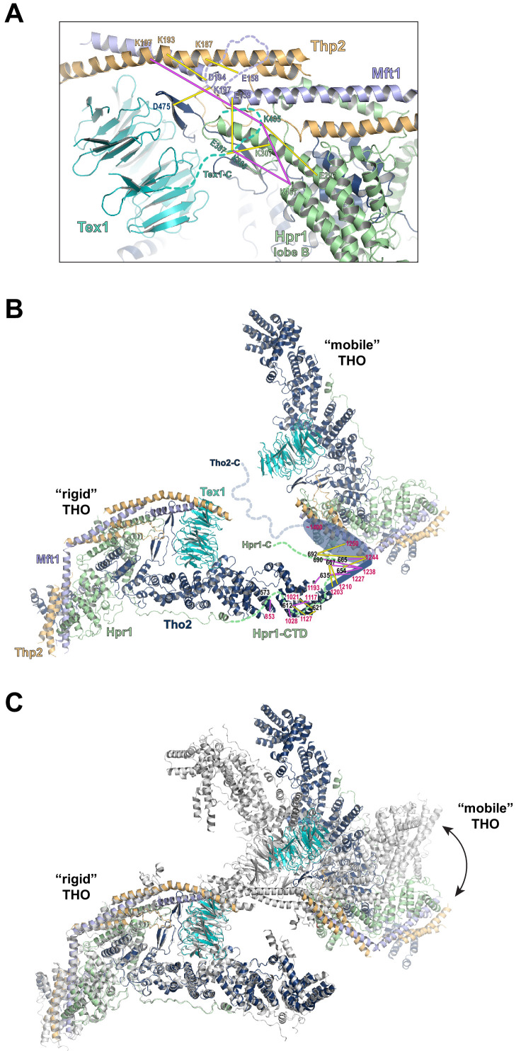
Figure 4—figure supplement 2. XL-MS data indicate the arrangement of the C-termini of Tex1 and Hpr1.

Figure 4—figure supplement 2.

(A) The C-terminal tail of Tex1 (residues 367–422) is localized near Hpr1 lobe B. The cross-linking sites are indicated by yellow lines. (B) The Hpr1-CTD binds to the C-terminus of the Tho2 ‘trunk’ and is localized close to the neighboring THO molecule. (C) Structural alignment of the dimeric THO assembly between our cryo-EM structure (colored as in B) and the recently reported THO–Sub2 structure (colored in gray, PDB ID 7AQO). Sub2 is omitted for clarity. The structures are aligned using one THO molecule, revealing significant flexibility in the relative orientation between the two THO molecules.
Figure 4—figure supplement 3. Gbp2 does not interact with Sub2.

Figure 4—figure supplement 3.

In vitro GST-pull down with excessive amount of Sub2 shows that Gbp2 does not make direct contact with Sub2 and Gbp2 still binds to THO•Sub2 under this condition.