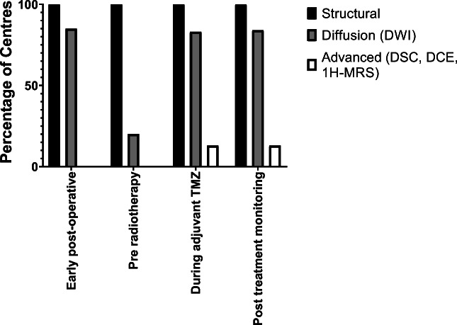
Fig. 2.
Imaging protocols at the different time points. Where the standardised protocol was described, most centres used diffusion weighted imaging (DWI), apart from at the pre-radiotherapy time point. Note that the figure shows the percentage of centres that routinely used advanced imaging techniques, as opposed to centres that only used advanced imaging techniques in selected patients, Structural = pre- and post-contrast T1-weighted, T2-weighted, FLAIR; DWI = diffusion weighted imaging; DSC = dynamic susceptibility contrast-enhanced MRI (perfusion); DCE = dynamic contrast enhanced MRI (permeability); 1H-MRS = 1H-MR spectroscopy

