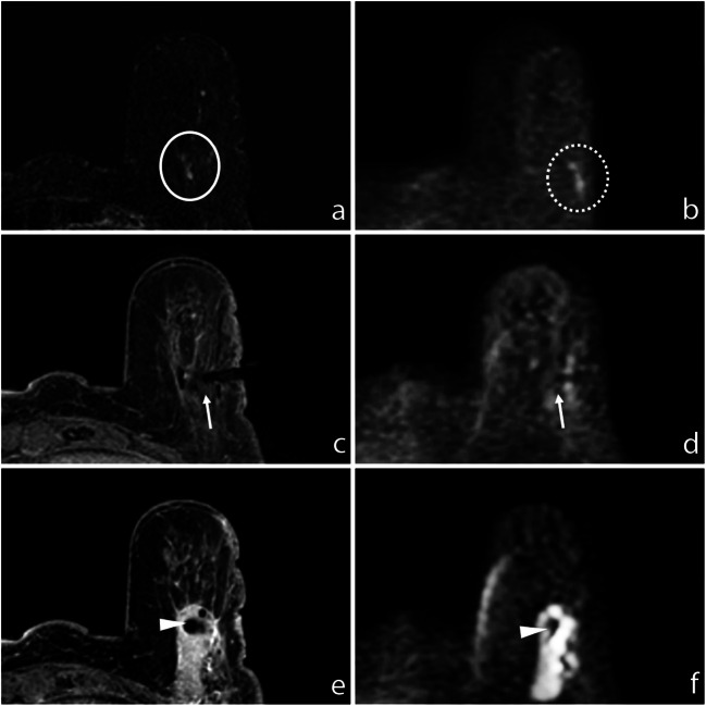
Fig. 5.
a Subtracted DCE 3D-THRIVE showing a non-mass lesion in the inferior sagittal plane of the left breast of a 71-year-old woman (circle). b DWI (b = 1300 s/mm2) showing hyperintensity of the enhancing lesion, slightly lateral-sided than real location due to spatial distortion artefacts (dotted circle). c T1-WI FS GRE showing the proper position of the biopsy device (arrow); the end of needle is well recognizable at (d) DWI (b = 1300 s/mm2, arrow). e T1-WI FS GRE showing the correct deployment of the site marker (arrowhead) that is also recognizable at (f) final DWI (b = 1300 s/mm2, arrowhead)

