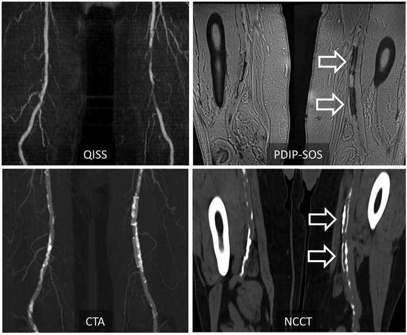
Figure 1 –
Representative case in a 72-year-old man with peripheral artery disease at 1.5T. PDIP-SOS MRI shows low-signal calcification involving the right superficial femoral artery, as well as two intra-vascular stents in the left superficial femoral artery (arrows). Corresponding non-contrast CT and contrast enhanced CTA shows good visual correlation in the location of vascular calcification between the two modalities. Calcification, however, appears to be more extensive on CT, possibly due to blooming artifacts. QISS MRA is also shown for comparison’s sake. PDIP-SOS, proton density weighted, in-phase 3D stack-of-stars; QISS, quiescent interval slice-selective

