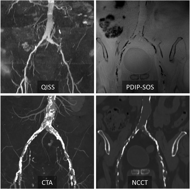
Figure 2 –
Representative case in a 77-year-old man with peripheral artery disease at 3T. Corresponding QISS MRA and CTA show peripheral artery disease in the common iliac arteries. PDIP-SOS MRI indicates extensive low-signal calcification involving the distal aorta, bilateral common and external iliac arteries. There is excellent correspondence in the location and extent of calcification between PDIP-SOS MRI and non-contrast CT (NCCT). PDIP-SOS, proton density weighted, in-phase 3D stack-of-stars; QISS, quiescent interval slice-selective

