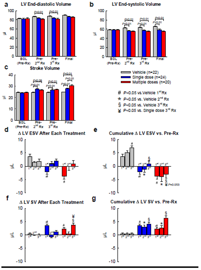
Figure 2. Echocardiographic assessment of LV volumes.

Echocardiographic studies were performed before each treatment and at the end of the study. a, LV end-diastolic volume; b, LV end-systolic volume; c, LV stroke volume; d, Changes in LV ESV after the 1st, 2nd, and 3rd treatments compared with the pre-treatment values; e, Cumulative changes in LV ESV after the 1st, 2nd, and 3rd treatments compared with the values measured before the 1st treatment; f, Changes in LV SV after the 1st, 2nd, and 3rd treatments compared with the pre-treatment values; g, Cumulative changes in LV SV after the 1st, 2nd, and 3rd treatments compared with the values measured before the 1st treatment. Data are means ± SEM.
