
Figure 4. Hemodynamic assessment of LV function.
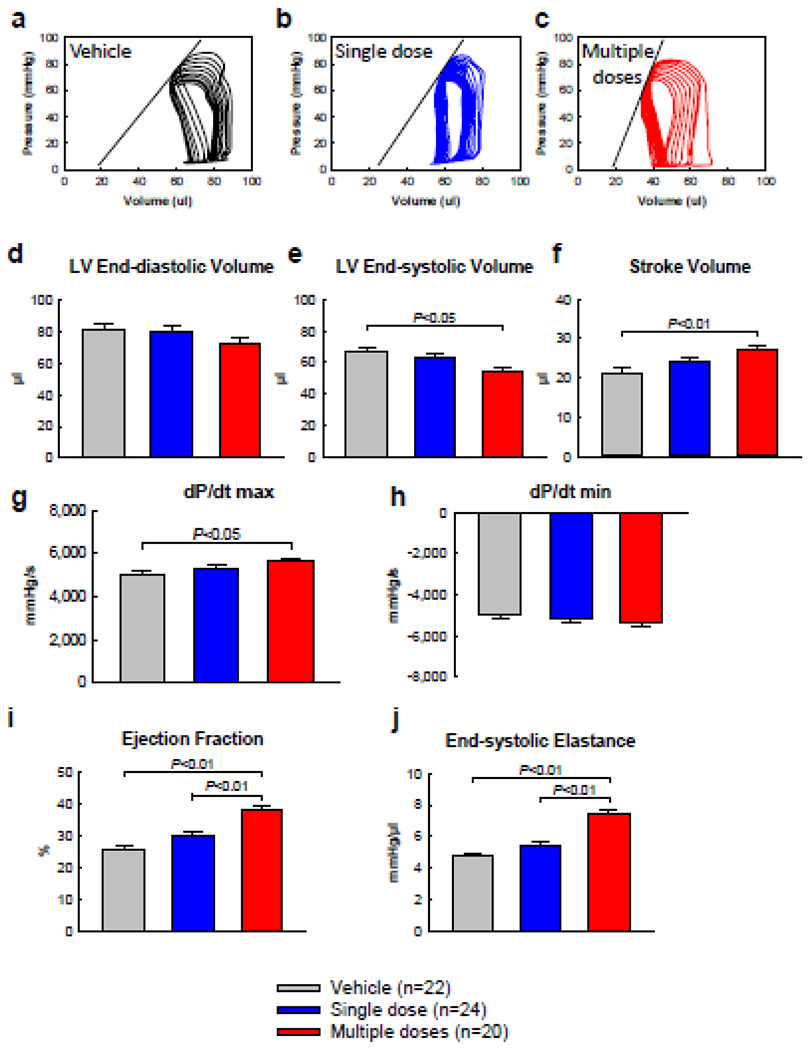
Invasive hemodynamic studies were performed with a 1F Millar conductance catheter 5 weeks after the 3rd treatment, just before euthanasia, a through c, Representative pressure-volume loops recorded during preload manipulation by brief inferior vena cava occlusions, d, LV end-diastolic volume, e, LV end-systolic volume, f, LV stroke volume, g, LV pressure dP/dt miximum and dP/dt minimum, h, LV EF. i, LV end-systolic elastance. Data are means ± SEM.