
Figure 1.
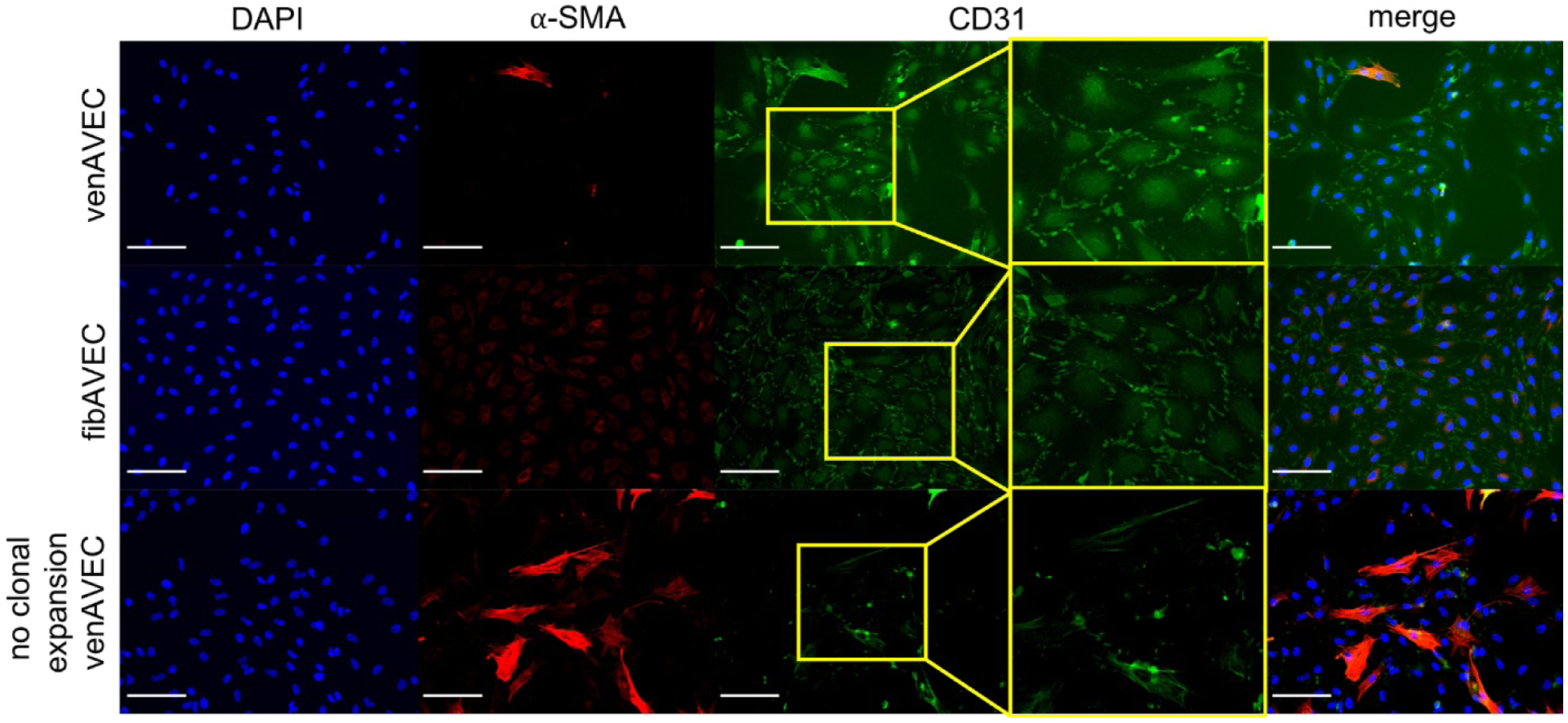
Both clonally expanded AVEC lines (venAVEC and fibAVEC) were stained using CD31 as an endothelial marker and α-SMA as a fibroblast marker, confirming endothelial phenotype. A line of non-clonally expanded venAVECs was used as a control. Scale bar is 100 μm.